
Pacing made visible
✓ Virtual Body Surface Map consisting of 14.000 leads
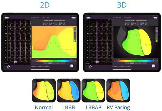
✓ 3D Imaging
✓ Beat-to-beat real time visual feedback while pacing
✓ Only 4 electrodes on the patient’s body
✓ Non-invasive
✓ Designed to be used intraoperative
✓ Intuitive app giving the operator full control
CardioSecur Vector-ECG imaging marks a new step in the evolution of ECG technology. It enables Electrophysiologists to get the full picture of the activation time of the patient’s heart mid procedure.
CardioSecur Vector-ECGi technology uses established principles to generate not only 12 or 22, but a total of 14.000 leads. In effect, this multitude of leads each act as dot, or pixel on a virtual 2D map of the heart’s electrical activity as seen from the surface of the patient’s body. We call this the Virtual Body Surface Map.
Vector-ECGi is non-invasive, runs on iOS iPads and uses only 4 electrodes. This makes V-ECGi suited to be employed during pacing procedures and allows for an unobstructed x-ray view of the patient's thorax. The operator can visually assess the electrophysiological condition of the heart and control beat-by-beat implantation positioning and outcome intraoperative with unparalleled precision.
One step further - 3D imaging
The next step in the development of the CardioSecur Vector-ECGi is the projection of the 2D Virtual Body Surface Map on a 3D anatomical model of the heart. The model will allow personalization by uploading a historic CT scan of the patient’s heart into the app. Continuous patient x-ray exposure is mitigated as the system links the heart’s morphology with its electrophysiologic activity, offering data insight previously unknown to practitioners and scientific research. In addition, the system displays ST elevations on the heart‘s surface. It reveals infarction areas and their spread in detail, also when the classical 12-lead ECG cannot show ST elevations.
*CardioSecur Vector-ECGi is in development and its approval pending. A beta-version is available to selected clinics for scientific research.